Hip Nail Failure Case
This case involves an elderly female who fell at home and fractured her hip. She was evaluated with x-rays and found to have an intertrochanteric hip fracture. Here are the initial x-rays:
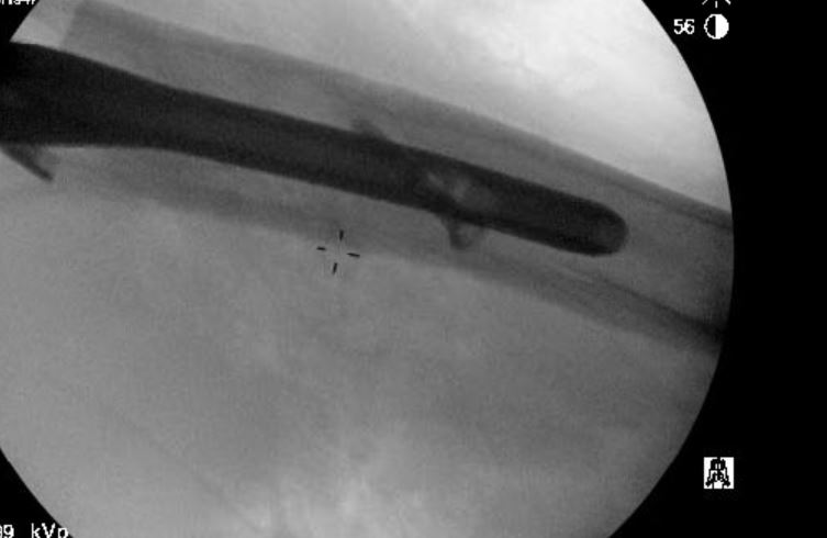
I consider this fracture pattern to be unstable due to the reverse obliquity of the fracture line. The patient was optimized and underwent intramedullary fixation with a cephalomedullary device (Zimmer Natural Nail). Here are the intra-operative fluoroscopy images.
As seen above, the cephalomedullary nail is in excellent position and the fracture is well reduced and aligned. Here are x-rays taken at routine follow up evaluations:
Here are x-rays at a later date:
You can see evidence of screw migration and fixation failure. At this point the patient had severe symptoms. A CT of the hip was obtained. Here are the coronal and sagittal reconstructions:
As seen, the hip screw has penetrated the femoral head and failed. There is a nonunion of the fracture as well. This patient required additional surgical treatment. Removal of the hip nail and placement of a partial hip replacement or hemiarthroplasty was recommended. Revision fixation was not considered advisable due to damage to the head and high risk of failure.
Here are the final x-rays showing the long stemmed partial hip replacement. The Zimmer/Biomet Arcos Modular Femoral Revision system was used.
The patient recovered well from the surgery and has regained functional abilities. I have noticed a higher incidence of these kinds of failures over the last few years. I suspect it is related to an aging population with more severe osteoporosis. Nevertheless, I think there is room for improvement in implant design to improve the success rate.


