Failure of Cementless Knee Replacement
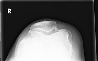
This case concerns a 63 year old male with advanced osteoarthritis of his right knee. He underwent a right cementless, posterior stabilized total knee replacement using Zimmer Nexgen LPS flex porous femoral component with trabecular metal standard primary patella and posterior stabilized trabecular metal monoblock tibial component. The knee replacement surgery was performed 8 years ago. The components were chosen due to provide the highest level of durability given patient's young age and high activity level. Cementless technology has the potential to be more durable than cementless implantation of joint replacement. Here are the initial x-rays:
 |
| Weight bearing x-rays of right knee revealing advanced varus arthritis |
 |
| x-ray of patella |
 |
| Immediate post-op x-rays showing successful implantation of cementless total knee replacement |
The patient returned 8 years later with recent onset of severe pain and difficulty moving his knee. Repeat x-rays were obtained:
 |
| Current x-rays show no obvious signs of failure |
MRI with metal artifact reduction sequences was obtained as well with no abnormalities noted.
He was taken to the operating room for arthroscopic evaluation and possible revision. The patella was found to be damaged upon arthroscopic evaluation so a decision was made to open up the knee and revise the components. Here are the arthroscopic pictures:
 |
| Arthroscopic pictures of patella showing damage to the polyethylene surface |
The components were all found to be well fixated to the bone. The femoral component was retained. Here pictures of the explanted components.
 |
| Explanted materials from revision knee replacement. The tibial post was broken off and there was severe damage to the polyethylene patella component articular surface |
Here are the post-operative x-rays from the revision showing a new patella component and stemmed LCCK tibial component. Note the metallic debris from cutting through the trabecular metal patella and tibial posts. Also note the screw down tibial polyethylene component which is a feature of the LCCK system. Hybrid fixation of the tibia was used with cemented metaphysis and cementless diaphysis. The splined cementless stem engages the cortices of the bone in the midshaft region.
 |
| Post-op knee x-ray |
 |
| Post-op knee x-ray |
 |
| Post-op knee x-ray |
Conclusion:
It seems that the polyethylene tibial post broke off due to cyclic loading caused by daily activities and low impact exercise activities that the patient participated in. This likely resulted in excessive wearing of the patella component as well with near wear through of the metal backed patella component.
There is much room for improvement in designing knee implants for the active adult <60 years of age. Present technology has a significant risk failure in < 10 years as seen in this case.Case presented with permission from the patient.
No comments:
Post a Comment